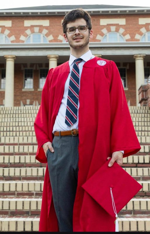
Tell me a little about yourself!
Hi, my name is Brendan Roess, and I graduated from NC State in 2020 with a B.S. in Biological Sciences. I am currently working in Dr. Nascone-Yoder’s lab during my gap year, and I am hoping to attend medical school in the near future. In my free time, I enjoy working out, going on runs, and spending time with family and friends.

I have been using the Zeiss Xradia 510 Versa X-Ray Tomography System. While this system is commonly used to scan non-biological samples, it has been extremely useful in scanning Xenopus laevis embryos. I like this instrument because of its relatively quick processing time and the high-resolution 2D projections it generates.
What have you been researching and how is it impacting the community?
Vertebrate anatomy has a characteristic left-right asymmetry in multiple organ systems. For example, your liver is on the right side of your body while your heart deviates slightly to the left side of your body. When this asymmetry is disrupted, it often results in birth defects that require immediate surgery to resolve. Xradia processing of X. laevis samples and the subsequent quantitative analysis of the samples will be instrumental in creating a model for stomach development in X. laevis. The stomach has its own left-right asymmetry, and in X. laevis, the stomach has a leftward curvature. The stomach model will have predictive qualities and be useful in mapping stomach development when specific biological pathways are perturbed. So far, several samples have been processed and are undergoing segmentation, which is necessary for future quantitative analysis. The instruments provided at the AIF are perfect for this type of project because they allow for rapid, overnight processing of samples. Additionally, the Zeiss Xradia X-Ray System generates 2D projections with almost cellular-level resolution.
The Xradia System has saved me many hours that would have been spent processing samples for IHC, imaging sections on a slide, and then rendering a 3D structure from those images using image processing software.
What have you learned from your experience at AIF?
The number one thing I have learned from my experience at the AIF is how important it is to take advantage of the wealth of tools available to you at NC State. If there is something at NC State that you think may be useful in your research, odds are you can use it, and a colleague is willing to show you how.
Xenopus laevis embryo
Best thing about AIF in 5 words or less?
The amazing staff!
Is there a staff member at AIF that has helped you?
Dr. Anton Jansson was extremely helpful and knew so much about the Xradia System at the AIF. He was generous with his time and always willing to meet via Zoom to answer questions regarding the image processing software Dragonfly. I cannot thank him enough!