Variable Pressure Scanning Electron Microscope – Hitachi SU3900
The Hitachi SU3900 variable pressure scanning electron microscope (VPSEM) is a conventional high resolution thermionic SEM, which allows the operator to control the specimen chamber vacuum level.
For conductive samples, the instrument is typically operated in high vacuum mode and images are collected with the Everhart-Thornley secondary electron detector. For insulating samples, the instrument is typically operated in variable pressure mode and images are collected with the Robinson backscattered electron detector. When operating in variable pressure mode, the chamber is back filled with a gas in order to allow imaging with little or no charging of the sample. Typically, the back fill gas is either N2 or He, but any non-corrosive, non-reactive gas can be used. Elemental analysis using the energy dispersive X-ray detector can be done in either high vacuum or variable pressure mode.
The Hitachi SU3900N SEM is equipped with the standard Everhard-Thornley secondary electron detector, a five segment solid state backscattered electron detector, Hitachi’s proprietary ultra-variable detector (gives a secondary electron image in variable pressure mode), an Oxford energy dispersive X-ray spectrometer, and a GW specimen current meter.
Solid state (5-segment) backscattered electron detector
Ultra-variable detector (UVD)
Oxford energy dispersive X-ray spectrometer
GW Specimen current meter
Capabilities
Backscattered electron images (atomic number contrast, configurable for topographic contrast)
Elemental analysis (X-ray spectra)
Elemental mapping (X-ray mapping)
Other standard and non-standard SEM techniques such as EBIC are possible
Accelerating Voltage
0.3 – 30 kV
Magnification
20X – 300,000X
Resolution (SE)
3.0 nm @ 30kV
15 nm @ 1 keV
Both are in high vacuum mode using the secondary electron detector
Resolution (BSE)
4.0 nm @ 30kV in variable pressure mode using the backscattered electron detector
Working Distance
3-60 mm
Specimen Stage
Five axis motor drive
Movable range
Rotate 360º
X = 150 mm
Y = 150 mm
Z (WD) = 5 to 85 mm
Eucentric tilt -20º to +90º
Observable sample area = 203 mm diameter
Maximum sample diameter = 300 mm
Maximum sample height = 130 mm
Maximum sample weight = 5 kg
Specimen Considerations
Accepts most samples: Conductors, Semiconductors, and Insulators can be observed
Including, but not limited to metals, semiconductor devices, ceramics, biological, polymer, textile, pharmaceuticals, even food, etc.
Sand Dollar (found at an NC beach) observed with the Robinson BSE detector at 500X.
Dendritic growth of Ag from a colloidal suspension observed with the Everhart-Thornley SE detector.
Etched Pearlite. Sample courtesy Lew Reynolds.
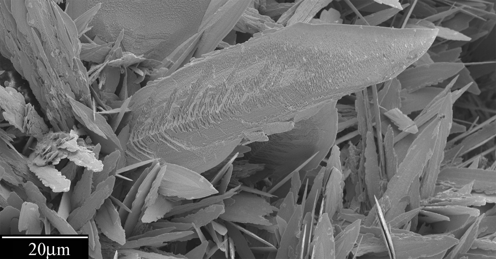
Pyrite (FeS) “framboids” filling the shell of a foraminifera in sediments from the continental shelf seaward of the North Island, New Zealand. Pyrite forms under reducing conditions in shallowly buried, organic-rich marine sediments. From the work of Lonnie Leithold.
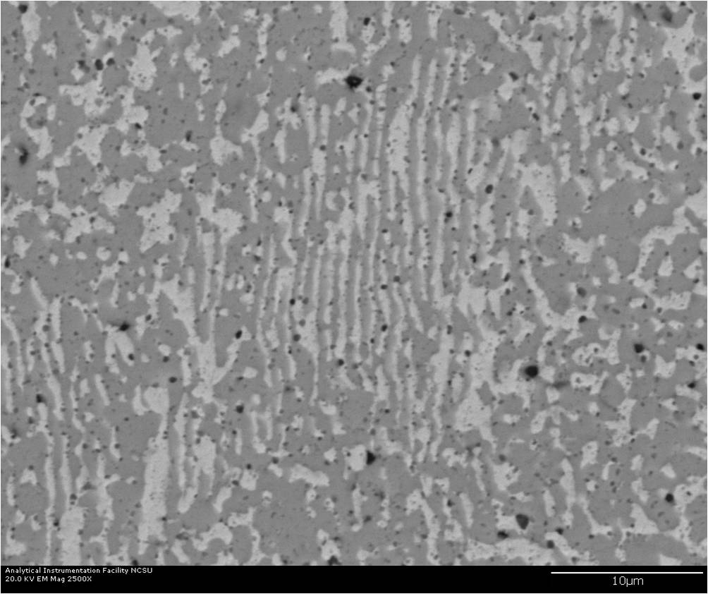
Lead-tin eutectic imaged with Robinson BSE detector. The BSE detector is sensitive primarily to changes in atomic number with higher atomic number materials having more contrast. In this case, the Pb is brightest and the Sn is darker. The dark particles observed are polishing media that are embedded in the sample. Sample courtesy Lew Reynolds.
Pollen sample taken from AIF staff member Chuck Mooney’s car.