|2024, AIF, Feature, NC State, Recent News, Students, think and do, User Spotlight
Meet April Sharp
By Anna Lumpkin
My name is April Sharp, and I am a third year PhD student in the Department of Applied Ecology, advised by Dr. Rebecca Irwin. I study disease dynamics of a protozoan gut parasite of bumble bees. When I’m not in the lab I love to draw and paint in addition to exploring the local parks and museums in the Triangle area. Beyond my degree I aspire to pursue a career in disease ecology research.
What instruments are you using for your research and why do you like them?
I have been using the Hitachi SU3900 Variable Pressure Scanning Electron Microscope to image bumble bee gut segments. This has been my first experience using SEM and it has been so exciting to see how much detail is captured in the images!
What have you been researching?
I study the role of pollen diet in shaping infection and transmission outcomes of a fecal-orally spread gut parasite, Crithidia bombi. This prevalent parasite infects bumble bees and is also found to spillover into solitary bee populations. Some pollen diets have been shown to significantly decrease gut parasite loads in the common eastern bumble bee, but the mechanism behind this medicinal effect is not fully understood. One factor that appears to play an important role in this effect is pollen morphology. The parasite-reducing effect has been found to be associated with pollen grains that have a distinctly spiked structure; however, through previous experiments, I have found that parasites in the feces of bees fed spiked pollen still go on to readily infect other bees. At the AIF I am using SEM to image the gut epithelial tissues of bumble bees fed spiked versus smooth pollen types to look for differences in tissue appearance. Higher instances of tissue abrasion or increased presence of mucins may indicate that spiked pollen upregulates immune responses which contribute to reduced parasite infections.
Pathogens play an important role in bee population declines, and additional stressors such as habitat loss and heat stress further exacerbate the negative effects of pathogens. The goal of my research is to better understand the role of pollen structure in mediating gut parasite infections in bumble bees. These findings could help inform management strategies to help limit the acquisition and infection intensities of gut parasites afflicting wild and managed bees.
What have you learned from your experience at AIF?
I have learned that no textbook diagram can really compare to the value of taking an up-close look at one’s study system! Getting to take detailed images of the gut tissues and anatomy of bees was an incredible learning experience. I also learned to always take advantage of the amazing research facilities at NC State.
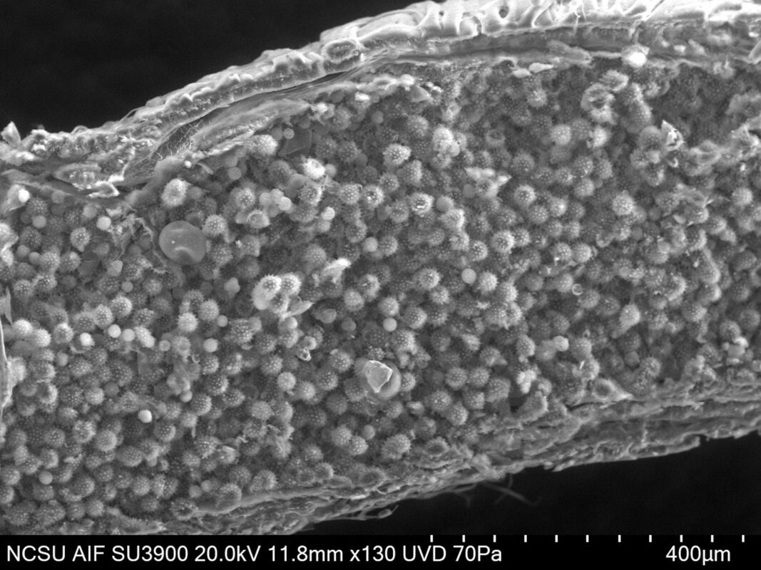
At the AIF I am using SEM to image the gut epithelial tissues of bumble bees fed spiked versus smooth pollen types to look for differences in tissue appearance.
This image shows the inside of a bumble bee’s midgut, full of sunflower pollen.
Best thing about AIF in 5 words or less?
Friendly experts with advanced instruments!
Is there a staff member at AIF that has helped you?
Thank you to Dr. Aaron Bell for his help with sample preparation and imaging. His expertise on microscopy techniques and protozoa has been a huge help to this project.